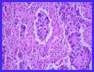
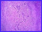
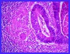
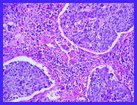
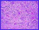
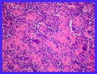
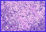
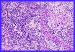
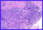
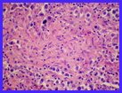
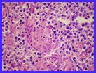
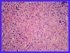
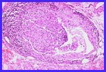
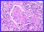
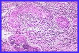
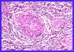
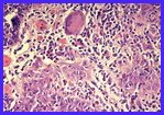
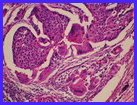
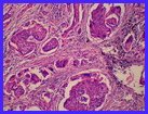
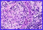
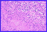
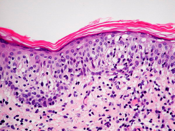
Granulomas may be encountered in association with a variety of malignant neoplasms in the following circumstances:
- Commonly in Hodgkin disease and non-Hodgkin T cell lymphomas, seminoma of the testis and ovarian dysgerminoma. Some individuals with seminoma may develop granulomatous lymphadenitis in non-metastatic lymph nodes in sites distant from the primary tumor. In Hodgkin disease granulomas may be seen in iiver and spleen which exhibit no evidence of Hodgkin disease involvement.
- Uncommonly in carcinomas arising from varied sites and in lymph nodes draining the site of carcinoma.
- Granulomatous reaction to a therapeutic agent; i.e. BCG, Interferon, Methotrexate.
- Coincidental occurrence of a systemic granulomatous disease and a malignant neoplasm.
The following images are examples of granulomas associated with malignant neoplasms
GRANULOMAS ASSOCIATED WITH MALIGNANT NEOPLASMS
Click on thumbnail image to view full-size image
1A- Hodgkin disease, lymph node; granuloma
1B- Hodgkin disease, lymph node; minute epithelioid granulomas
1C- Hodgkin disease, lymph node; minute epithelioid granulomas
2A- Non-Hodgkin lymphoma, lymph node
2B- Non-Hodgkin lymphoma, lymph node
4A- Seminoma-non-necrotizing granulomas
3B- Non-Hodgkin lymphoma, skin
3C- Non-Hodgkin lymphoma, skin
4D- Seminoma-early necrosis in granuloma with apoptotic bodies
4E- Seminoma-necrotizing granuloma
4F- Seminoma-necrotizing granuloma
4B- Seminoma-non-necrotizing granuloma
4C- Seminoma-non-necrotizing granuloma
4-G Seminoma-intratubular granulomas
4H- Seminoma-intratubular granuloma
4 I- Seminoma-granulomatous angiitis
4J- Seminoma-granulomatous angiitis
5- Adenocarcinoma of colon metastatic to lymph node
6A- Adenocarcinoma of colon, poorly differentiated metastatic to lymph node
6B- Adenocarcinoma of colon, poorly differentiated metastatic to lymph node
7- Adenocarcinoma of stomach, poorly differentiated metastatic to lymph node
8- Large cell carcinoma of lung
9A- Squamous cell carcinoma of lung
9B- Squamous cell carcinoma of lung
11- Squamous cell carcinoma, poorly differentiated, urinary bladder
12A- Melanoma following BCG therapy
12B- Soft tissue around melanoma following BCG therapy
12C- Granulomatous angiitis at site of intralesional BCG therapy for melanoma
12D- Granulomatous angiitis at site of intralesional BCG therapy for melanoma
12E- Lymph node draining site of intralesional BCG therapy for melanoma
12F- Lymph node draining site of intralesional BCG therapy for melanoma
10A- Squamous cell carcinoma of lung
10B- Squamous cell carcinoma of lung
10C- Mediastinal lymph node draining squamous cell carcinoma of lung
3A- Non-Hodgkin lymphoma, skin
13- Invasive duct carcinoma of breast
14A- Mycosis fungoides, skin,low magnification (courtesy of Dr. Emmanuel Maicas-with permission)
14B- Mycosis fungoides, skin,med. magnification (courtesy of Dr. Emmanuel Maicas-with permission)
14C- Mycosis fungoides, skin, granulomatous,high magnification (courtesy of Dr. Emmanuel Maicas-with permission)
