Microscopic Images of Lung Involvement in Sarcoidosis
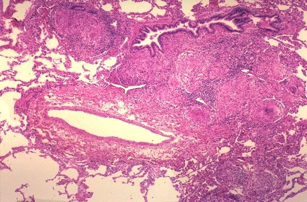
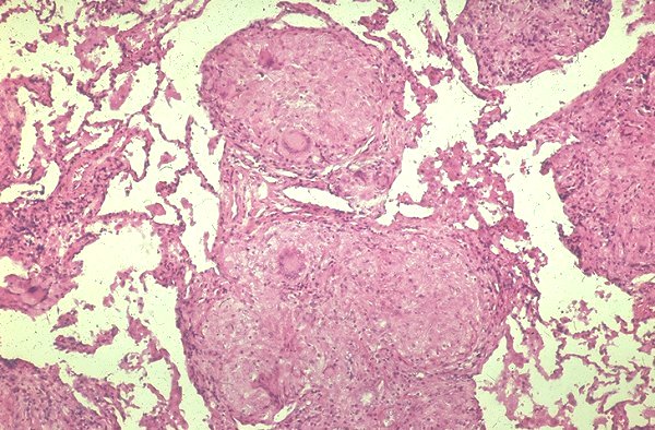
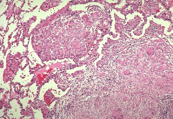
The location and appearances of granulomas in the lungs in sarcoidosis are varied. A pattern that is frequently seen in open lung biopsies and in autopsies is the localization of granulomas around airways, blood vessels,(Fig. 1) and fibrous septae (Fig. 2). Lymphatics are located in these sites and it is thought that this pattern is the result of dissemination of the inciting agent(s) via the lymphatics. The finding of sarcoid granulomas in lymphatics and the involvement of intrathoracic lymph nodes in all patients with intrathoracic sarcoidosis supports this interpretation.
Fig. 1 Granulomas adjacent to bronchiole and pulmonary artery.
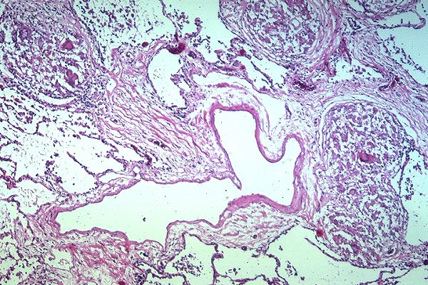
Fig. 2 Granulomas adjacent to fibrous septum. Note presence of pulmonary vein and lymphatic
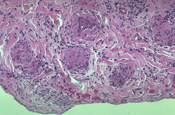
Granulomas may be discrete and located within alveolar walls (Figs. 3, 4)
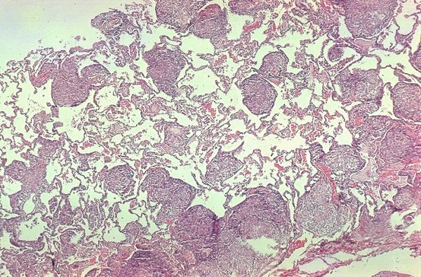
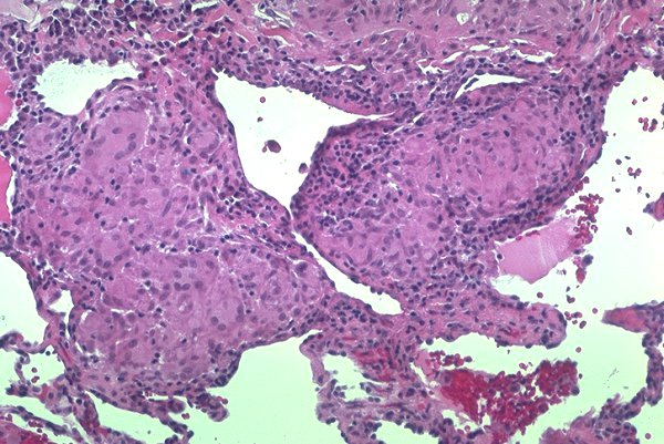
Granulomas may become confluent (Fig. 5) and form nodules of varying size (Fig. 6)
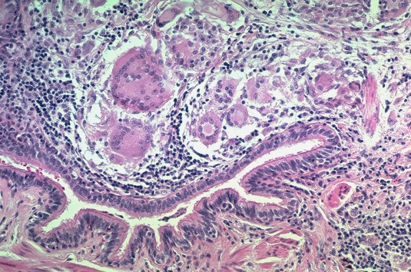
Granulomas often involve airways (Fig 7) and pleura (Fig. 8). Clinical manifestations of pleural involvement are rare.
Fig. 3 Discrete interstitial granulomas
Fig. 4 Interstitial granulomas
Fig. 5 Granulomas becoming confluent
Fig. 6 Confluent granulomas forming a nodule
Fig. 7 Granulomas involving a bronchiole
Fig. 8 Granulomas involving visceral pleura
Click on images to see a larger image