Necrotizing Sarcoid Granulomatosis (NSG)
Click on the following thumbnail images of a case of NSG to view larger images
Necrotizing sarcoid granulomatosis (NSG) is an uncommon condition that primarily involves the lungs; a small number of cases exhibiting extrapulmonary involvement have been reported. As of this writing (5/04) approximately 100 cases have been published. The overall prognosis for patients with NSG is excellent
NSG usually presents radiographically as multiple bilateral nodular lung nodules but may present as a solitary nodule. Evidence of cavitation and hilar lymphadenopathy are occasionally seen. Most patients experience pulmonary symptoms.
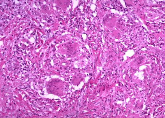
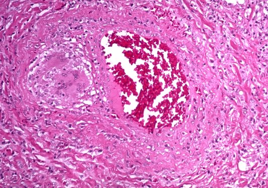
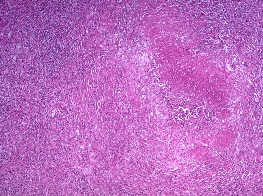
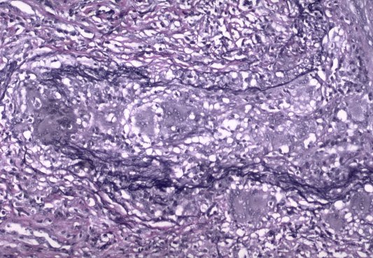
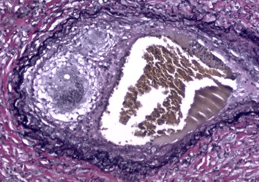
The characteristic pathologic features of NSG are confluent granulomas, granulomatous angiitis, and necrosis within granulomas varying from tiny punctuate foci to large foci of infarct-like necrosis. Granulomatous infection should be excluded before making a diagnosis of NSG.
Although the nature of NSG is unknown it appears likely that most cases represent “nodular sarcoidosis”. “Nodular sarcoidosis” is primarily a radiologic diagnosis, the pathologic features of which are not well-documented. Approximately 5% of cases of sarcoidosis present radiographically as “nodular sarcoidosis”. In a large series of open lung biopsies from patients with sarcoidosis the combination of confluent granulomas, granulomatous angiitis, and necrosis , the diagnostic features of NSG, was found in 5% of the specimens.


NSG:Chest X-ray. Single large nodule, RLL
NSG:Gross specimen photograph of excised nodule
NSG:Microscopic-Confluent granulomas
NSG:Microscopic-Confluent granulomas and necrosis
NSG:Microscopic-Granulomatous angiitis
NSG:Microscopic-Granulomatous angiitis, elastica stain
NSG:Microscopic-Granulomatous angiitis, elastica stain