GRANULOMATOUS PULMONARY ANGIITIS SARCOIDOSIS
Granulomatous pulmonary angiitis is a frequent finding in sarcoidosis. It may be seen in two-thirds of open lung biopsy specimens, even more frequently in autopsy lungs, and only occasionally in transbronchial biopsies.
Venous involvement is seen far more frequently than arterial involvement. Granulomas may also be seen in lymphatics (Fig. 14).
Pulmonary arterial aneurysms are not seen despite the occurrence of focal full-thickness elastic tissue destruction in some cases (Fig. 10). This is likely due to the low pressure in the pulmonary circulation.
Rare instances of pulmonary hypertension related to granulomatous pulmonary angiitis in sarcoidosis have been reported,
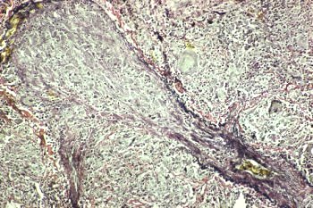
Elastic tissue stains are very helpful in identifying these lesions, particularly in small vessels where the lumen is completely obliterated (Figs. 3,4,5)
Granulomatous angiitis, although highly characteristic, is not specific for sarcoidosis and may also be seen in infectious granulomatous diseases, Wegener's granulomatosis, foreign body embolization, schistosomiasis, necrotizing sarcoid granulomatosis, beryllium disease, intralesional BCG injection, and seminoma.
 | ||||||
| ||||||
| ||||||
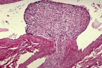
1. Polypoid intimal granuloma - pulmonary artery
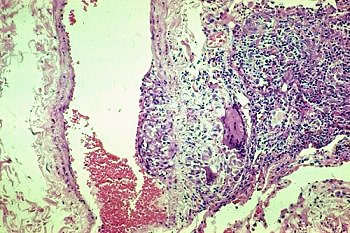
2. Transmural granuloma - pulmonary vein
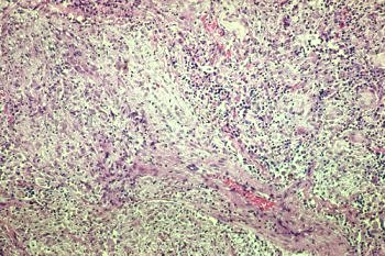
3. Granulomatous obliteration of small vein
4. Granulomatous obliteration of small vein - Elastic stain
Additional photos of granulomatous pulmonary angiitis
Focal complete elastica destruction-no aneurysm
From transbronchial biopsy-low magnification
From transbronchial biopsy-high magnification