LESIONS ASSOCIATED WITH PRIMARY TUBERCULOSIS: Initial infection with Mycobacterium. tuberculosis in an immunocompetent individual usually occurs in an upper region of the lung producing a sub-pleural lesion called a Ghon focus. Granulomatous involvement of peribronchial and/or hilar lymph nodes is frequent in primary tuberculosis due to lymphangitic spread from the Ghon focus. The early Ghon focus together with the lymph node lesion constitute the Ghon complex. These lesions undergo healing and over time usually evolve to fibrocalcific nodules. The combination of late fibrocalcific lesions of the lung and lymph node which evolved from the Ghon complex is referred to as the Ranke complex.
Fig. 3- Larger sub-pleural fibro-calcific nodule (healed Ghon focus)
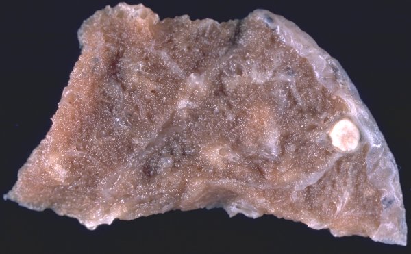
Fig. 2- Apical, sub-pleural fibro-calcific nodule (healed Ghon focus) Emphysema
Fig. 1-Sub-pleural fibro-calcific nodule (Healed Ghon focus)
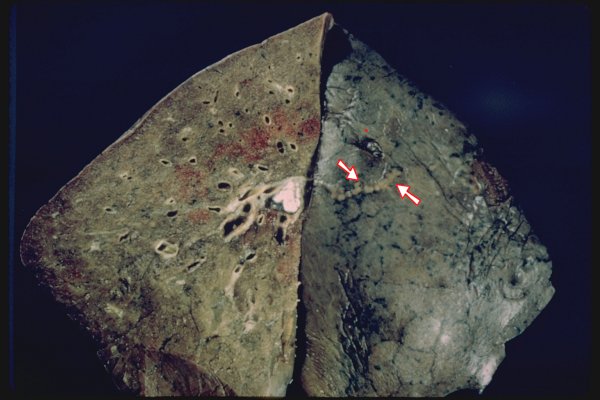
Fig. 5A- Ranke complex. Subpleural fibrocalcific nodule (white arrows) and thickened, scarred lymphatic vessel (red arrows)
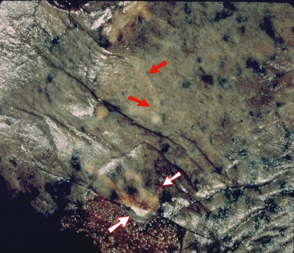
Fig. 5B- Ranke complex. Fibrocalcific lesion of peribronchial lymph node and thickened, scarred lymphatic vessel (arrows)
TUBERCULOMA: A well-circumscribed tuberculous lesion usually presenting as a "coin lesion" on chest X-ray . Usually a single nodule but may be more than one. These lesions are usually larger than the Ranke lesions and probably also represent a late phase of a primary tuberculous infection.
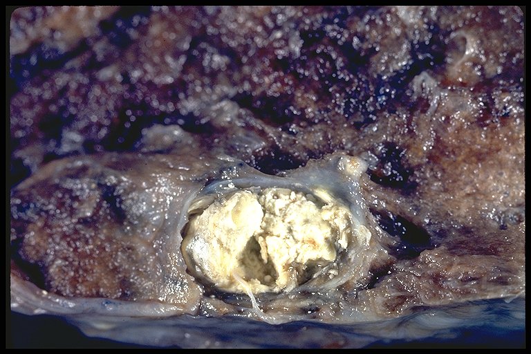
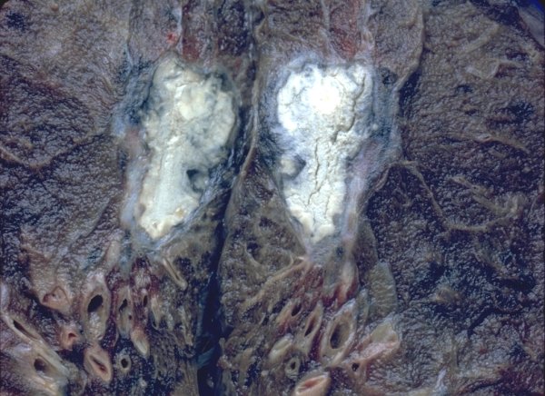
Sub-pleural tuberculoma.. Good example of "caseous" necrosis
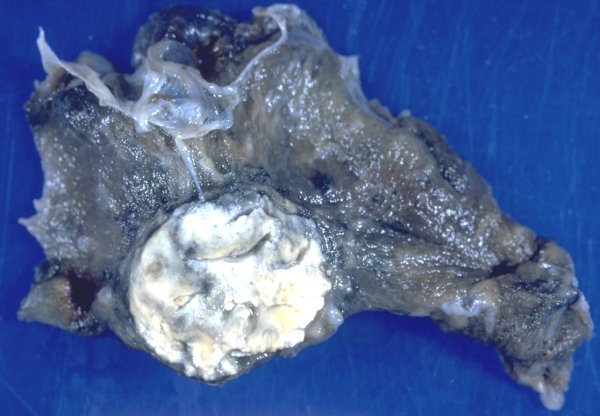
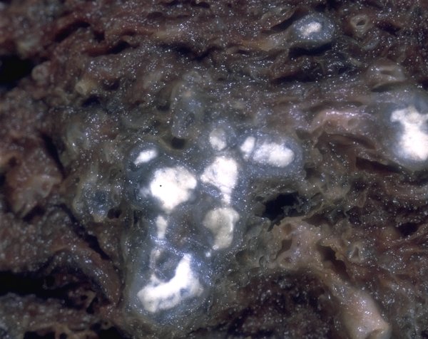
Sub-pleural fibrocalcific tuberculoma..
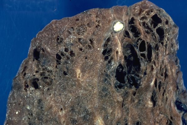
Fig. 4- Heavily calcified, healed focus of primary infection
Click on thumbnail image to view full-size image